Michigan Splint: intraoral scanner protocol for dental clinics
Learn how to create a Michigan splint with an intraoral scanner. Offer precise digital bruxism splints through a clinic-ready workflow.
Discover how intraoral scanner technology is transforming dental practices. In this article, we show you how to take precise, personalized scans, with a special focus on creating an occlusal splint, also known as a Michigan splint.
The bruxism splint is custom-made for each patient using the latest dental 3D printing technology and high-strength biocompatible materials. This process ensures a precise and comfortable solution for treating bruxism and other mandibular disorders, significantly improving the quality of dental care.
Step-by-step intraoral scanner protocol: optimize Michigan splint design
The intraoral scanning protocol for creating Michigan splints follows a clear, structured process. Below, we outline the detailed steps to achieve the best results in your clinic.
Patient preparation
Before starting the scanning process, it is essential to prepare both the patient and the equipment to ensure a successful and accurate scan:
- Prepare the patient: Make sure the patient is comfortable in the dental chair and that their mouth is clean and dry. This helps ensure an optimal scan without interference.
- Explain the procedure: Brief the patient on the process, explaining each step so they remain calm and cooperative during scanning.
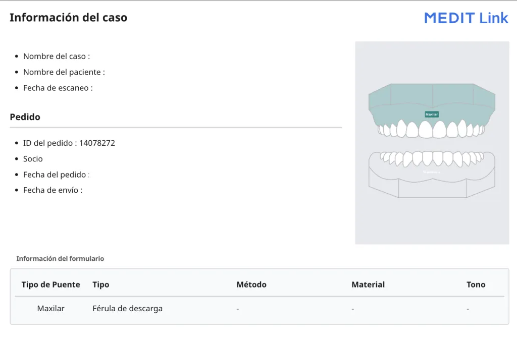
- Confirm the treatment details: Check the scan order, making sure it is correctly aligned with the planned treatment, whether for a Michigan splint or another appliance, and specify whether the scan is for the upper or lower arch.
- Prepare the intraoral scanner: Make sure the scanner is in perfect working order, fully charged, and fitted with clean scanning tips in optimal condition for accurate results.
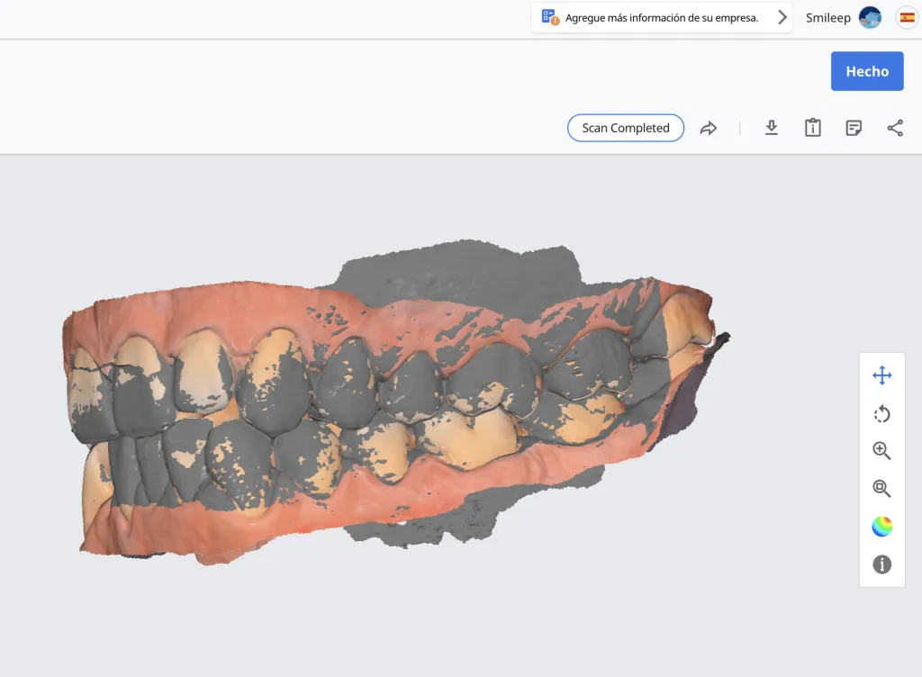
Maximum intercuspation occlusion scan (MIP) for the Michigan splint
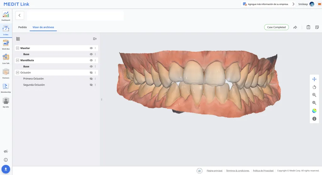
The maximum intercuspation occlusion scan (MIP) is essential for achieving a perfect fit for the Michigan splint. This position, in which the upper and lower teeth reach maximum contact during mandibular closure, is critical for designing custom occlusal splints and other dental appliances.
This precise and detailed scanning process is key to ensuring that the Michigan splint is manufactured with maximum accuracy, optimizing both comfort and treatment effectiveness.
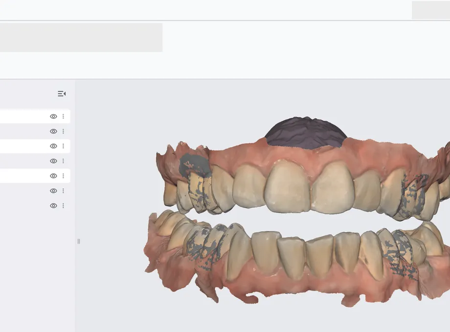
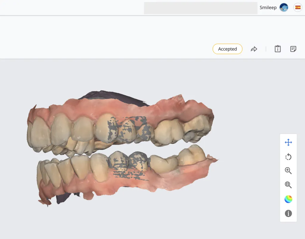
After fully scanning the upper and lower arches, it is necessary to perform the maximum intercuspation occlusion scan (MIP). To do this, capturing 2 to 3 posterior teeth is essential, allowing the scanner to align both bites accurately.
This step is crucial to guarantee a perfect fit for the Michigan splint or any other appliance being designed, ensuring the correct relationship between the arches.
This methodology significantly simplifies the scanning process, making it easier to adjust the vertical dimension precisely according to the designer’s assessment. That allows for more detailed customization in the design of the Michigan splint.
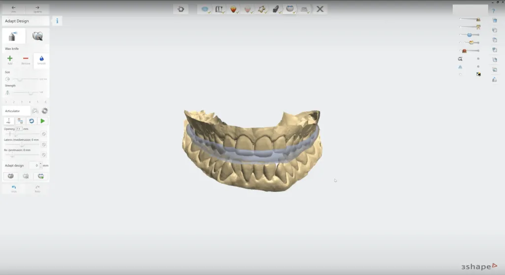
“The only consideration to keep in mind is that, in more complex bite cases, adjustments to the contact points may be required to ensure optimal results.”
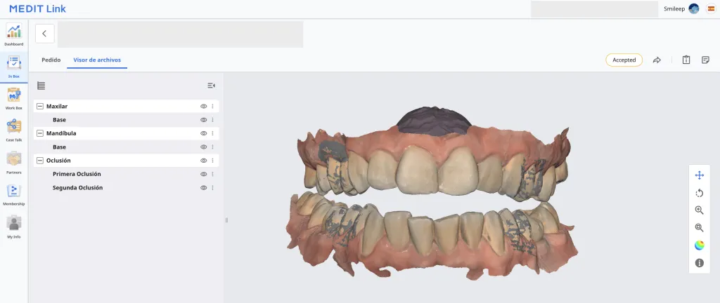
Occlusion scan with digital vertical dimension for the Michigan splint
Before starting the occlusion scan, it is crucial to prepare the patient properly to ensure an accurate fit for the Michigan splint. Below are the steps to follow for a successful scan, with particular attention to the vertical dimension:
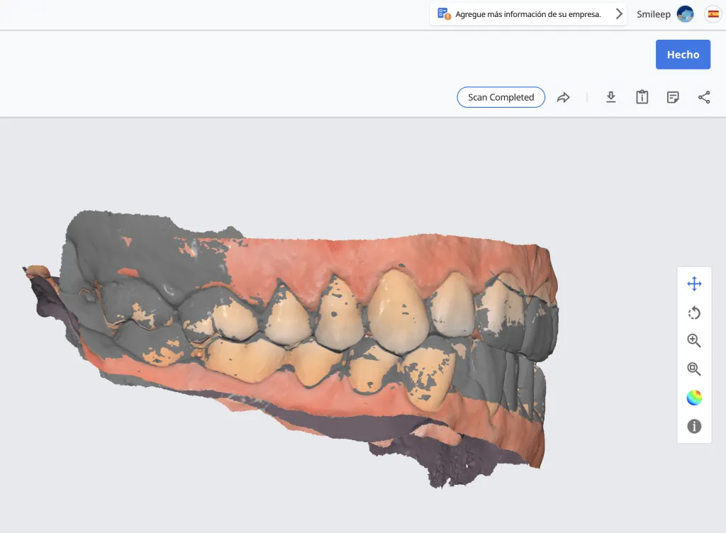
- Place the Lucia jig: Place a Lucia jig in the patient’s mouth to establish the desired vertical dimension, which is key to the proper fit of the splint.
- Patient comfort: Make sure the patient is comfortable and relaxed with the Lucia jig in place. Comfort during the process is essential for an accurate scan.
- Verify the vertical dimension: Check that the established vertical dimension is appropriate for the clinical case. An incorrect vertical dimension setting could affect the fit of the Michigan splint.
The Lucia jig is a commonly used device for establishing vertical dimension in the patient’s mouth, allowing a more precise bite adjustment. However, other alternatives also exist, such as bite waxes or Long strips, which may be used depending on the patient’s specific needs and the clinical case. It is important to select the most suitable option to achieve an optimal result in the design of the Michigan splint.
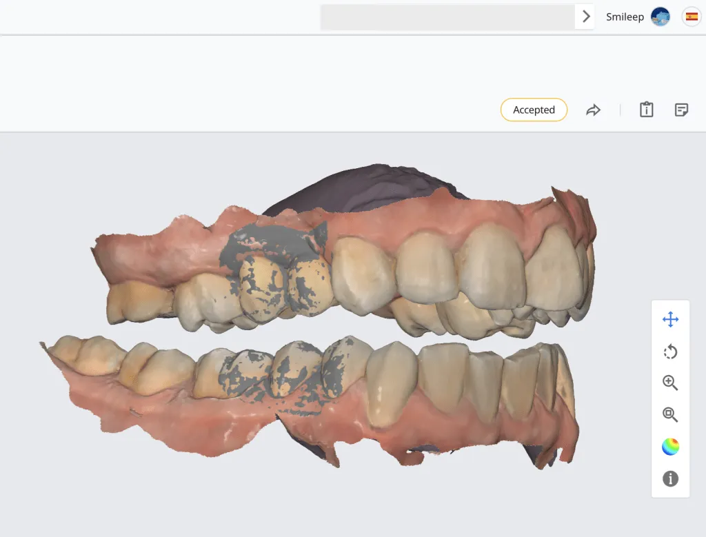
“Digital scanning of the vertical dimension allows us to design with greater precision and a better understanding of the patient’s occlusion, ensuring an optimal fit. This protocol not only improves results, but also minimizes unnecessary adjustments during chairside fitting.”
Add a Medit intraoral scanner to your clinic
The Medit intraoral scanner is an essential tool for transforming your dental practice, standing out for its advanced technology and its ability to deliver precise and efficient results. With a well-established reputation in digital dentistry, the Medit scanner optimizes the workflow and improves the patient experience, enabling faster diagnoses and more effective treatments.